This web page was produced as an assignment for Genetics 677, an undergraduate course at UW‐Madison.
Primary literature review: mutations that underlie the Tangier disease phenotype
Tangier disease caused by compound heterozygosity for ABCA1 mutaions R282X and Y1532C. Cameron J, Ranheim T, Halvorsen B, Kulseth M, Leren T, Berge K. Atherosclerosis 209 (2010) 163-166. doi:10.1016/j.atherosclerosis.2009.08.039
Introduction
Tangier disease is characterized by low blood plasma levels of HDL (high density lipoprotien) [1]. Low HDL levels significantly increase the risk of atherosclerosis, one of the leading causes of death in the United States and worldwide [2].
Because HDL production requires the activity of many genes, many different mutations can contribute to low levels of HDL [1]. To characterize the relationship between specific mutations and the Tangier disease phenotype, scientists examined the genotype of a forty-year old man with Tangier disease for three different genes involved in cholesterol metabolism: ABCA1, LCAT and apoA-1.
ABCA1 codes for ATP-binding cassette transporter A1, which is necessary for transporting cholesterol and lipids from inside cells to the bloodstream. LCAT encodes lecithin:cholesterol acyltransferase, which esterifies cholesterol so that it can properly form HDL particles. Finally, apoA-1 produces apolipoprotein A-1, which acts as a scaffold for cholesterols and lipids to form HDL particles.
Results
After sequencing the proband's DNA, the authors did not find mutations in LCAT or apoA-1.
However, they did find that the proband had two mutant copies of the ABCA1 gene, and that they were different mutations: R282X and Y1532C. The proband's mother and brother both carried one copy of R282X, and his father and daughter carried one copy of Y1532C.
The authors also studied the effect of the novel mutation Y1532C at the cellular level by examining HeLa cells transfected with the mutant genes. In cells carrying the Y1532C mutation, ABCA1 proteins failed to localize to the cell membrane (see figure at right).
Conclusions
The authors concluded that the low HDL concentration observed in the proband was caused occurred because ABCA1 could not reach the cell membrane to perform its transport functions. They hypothesized that the mutated site in the Y1532C allele (amino acid # 1532) causes this phenotype by weakening the ABCA1 protein's affinity for apoA-1, which helps ABCA1 localize to the cell membrane in healthy cells.
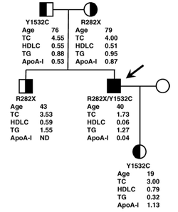
Figure 1 from Cameron et al.: phenotypes of the proband's family; the male proband is indicated by the arrow, carriers (who had lower than normal, but not harmfully low HDL levels) are shown as split circles (females) or squares (males)
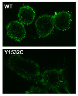
Column 1 from Figure 4: these confocal micrographs show that Y1532C ABCA1 proteins do not localize to the cell membrane like the WT -and thus cause reduced cholesterol efflux
Sources:
1. Tangier disease caused by compound heterozygosity for ABCA1 mutaions R282X and Y1532C. Cameron J, Ranheim T, Halvorsen B, Kulseth M, Leren T, Berge K. Atherosclerosis 209 (2010) 163-166. doi:10.1016/j.atherosclerosis.2009.08.039.
2. Cellular cholesterol trafficking and compartmentalization. Ikonen E. Nat Rev Mol Cell Biol. 9 (2008) 125-138.
